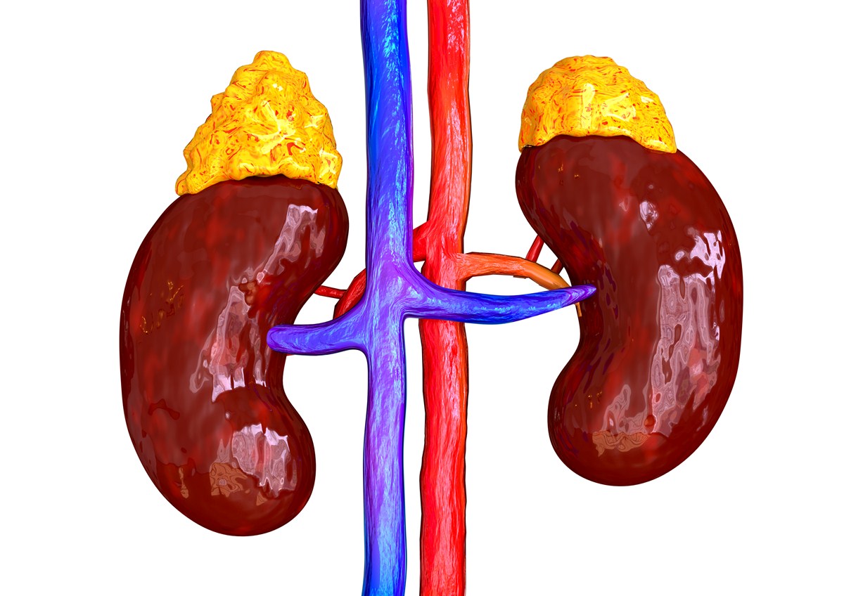


In addition, multiple profiles of unmyelinated nerve fibers were demonstrated in all treated specimens. These cells increased significantly in number, length of their telopodes, and secretory activity after melatonin treatment. The TCs were small branched cells scattered in the adrenal glands around cortical cells, chromaffin cells, nerve fibers, and blood vessels. Moreover, these SGC cells, Schwann cells, fibroblasts, and progenitor stem cells showed a stimulatory response. The most interesting feature in this study is the presence of small granule chromaffin cells (SGC) and telocytes (TCs) for the first time in the adrenal glands of sheep.

The ganglion cells of the melatonin-treated group showed a significant increase in diameter with numerous rER. Chromaffin cells in the control group expressed moderate immunoreactivity to Synaptophysin and tyrosine hydroxylase, compared with intensified expression after melatonin treatment. Piecemeal degranulation mode of secretion was recorded after melatonin treatment. Exocytosis of secretory granules to the lumen of blood vessels was evident in the treated group. The cytoplasm of these cells showed numerous mitochondria, rough endoplasmic reticulum (rER), Golgi apparatus, lysosomes, and glycogen granules. The most striking ultrastructural features in the medulla of the treated group were the engorgement of chromaffin cells with enlarged secretory granules enclosed within a significantly increased diameter of these cells. Our results revealed that the cells of adrenal cortex of the treated animals were separated by wide and numerous blood sinusoids and showed signs of increase steroidogenic activity, which are evidenced by functional hypertrophy with increase profiles of mitochondria, smooth endoplasmic reticulum, and lipid droplets. Adrenal glands of 15 Soay ram were examined to detect the effect of melatonin treatment. In this study, we attempted to investigate the effect of exogenous melatonin treatment on the adrenal cortex and medulla using several approaches. Studies on the melatonin effect on the adrenal glands which are important endocrine organs, controlling essential physiological functions, are still deficient. Endogenous melatonin is a hormone secreted by pineal gland it has several roles in metabolism, reproduction, and remarkable antioxidant properties.


 0 kommentar(er)
